Zebrafish Core Unit
Models for pediatric cancer
We support researchers to develop novel therapeutic strategies by leveraging the excellent imaging and drug-screening capabilities of zebrafish to unravel the etiology and disease-driving mechanisms of pediatric cancer.
Background
Cancer is a complex disease and is ideally studied in a natural environment as it is greatly influenced by cell interactions, but also by mechanical cues from the surrounding tissue.
The zebrafish is a vertebrate model organism gaining increasing attention in cancer research (https://doi.org/10.3389/fonc.2017.00186). Offering superior live imaging abilities, zebrafish larvae allow us to follow cancer cells within the intact organism and monitor interactions with other cell types in detail. Furthermore, zebrafish larvae are practical to carry out preclinical drug screens in vivo. We make use of these advantages and model pediatric cancer in zebrafish to better understand tumor onset and progression.
Approach
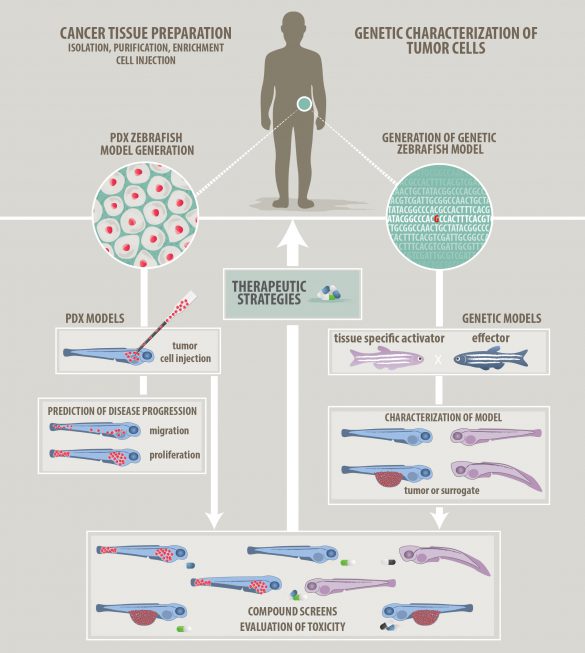
To model pediatric cancer we have two options, either genetic modelling by introducing oncogenes or a xenotransplantation strategy of human tumor cells (Figure 1). Our models allow us to observe tumor cell behavior down to the subcellular level by intravital microscopy and we can capture interactions with the tumor microenvironment.
Figure 1: Strategies to model cancer in zebrafish
Zebrafish models of pediatric sarcomas
Ewing sarcoma and osteosarcoma are the most frequent bone cancers found in children and young adolescents with dismal outcome, especially for patients with metastasis or after relapse. Here, zebrafish can be applied in innovative ways to tackle open questions like: What is the cell of origin in Ewing sarcoma? What are main drivers of metastasis in Osteosarcoma?
We have successfully established zebrafish xenograft models for Ewing sarcoma and Osteosarcoma (Figure 2). Our recent screen identified compounds and particular compound combinations, which are highly effective against Ewing sarcoma cells in our xenograft setting (see https://doi.org/10.1016/j.canlet.2022.216028 ).
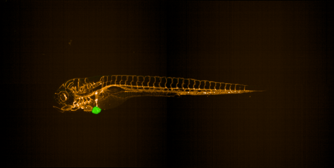
Figure 2: Zebrafish Ewing sarcoma xenograft
Zebrafish drug screening platform (ZANDR)
Particularly zebrafish larvae are well-suited to perform cost-effective screens for small molecules with therapeutic potential. The FFG funded Zebrafish platform Austria for preclinical drug screening (ZANDR; see www.zandr-ccri.at), was established at the Children´s Cancer Research Institute in 2019 and is a unique platform for phenotypic screening of small compounds in zebrafish models of human diseases. We anticipate that drug screening in zebrafish will provide valuable information to decide, which compounds to advance towards clinical application.
ZANDR is a zebrafish-optimized and automated screening platform which is open to the scientific community.
Please contact us if you are interested in collaboration.
We support technological development
We are striving to enhance the available tools and methods for zebrafish research with a current focus on
- Automating and improving the small compound screening workflow for zebrafish https://rdcu.be/fiHNJ; https://doi.org/10.1007/978-1-0716-4418-8_9
- Bringing new imaging modalities to zebrafish
- Generating reporter strains for tumor cell interactions in zebrafish
We offer the following services
- Xenografts of human cells into zebrafish larvae and drug screening
- Image acquisition and analysis of larvae or cells on a high-content imager
- Providing zebrafish eggs for microinjections
- Generation of zebrafish transgenic lines or CRISPR lines
- Generate genetic zebrafish tumor models using different cancer genes e.g. HRAS, KRAS, EWS-FLI1, tp53